Essential idea: Immunity is based on recognition of self and destruction of foreign material.
11.1 Antibody production and vaccination
UNDERSTANDINGS:
U11.1.1 Every organism has unique molecules on the surface of its cells.
U11.1.2 Pathogens can be species-specific although others can cross species barriers.
U11.1.3 B lymphocytes are activated by T lymphocytes in mammals.
U11.1.4 Activated B cells multiply to form clones of plasma cells and memory cells.
U11.1.5 Plasma cells secrete antibodies.
U11.1.6 Antibodies aid the destruction of pathogens.
U11.1.7 White cells release histamine in response to allergens.
U11.1.8 Histamines cause allergic symptoms.
Histamine is an important protein involved in many allergic reactions. Allergies are caused by an immune response to a normally innocuous substance (i.e. pollen, dust) that comes in contact with lymphocytes specific for that substance, or antigen. In many cases, the lymphocyte triggered to respond is a mast cell. For this response to occur, a free-floating IgE (an immunoglobulin associated with allergic response) molecule specific to the antigen must first be attached to cell surface receptors on mast cells. Antigen binding to the mast cell-attached IgE then triggers the mast cell to respond. This response often includes the release of histamine. It causes contraction of smooth muscle and dilation of capillaries.
Histamine causes many of the symptoms of allergies, such as a runny nose or sneezing. When a person is allergic to a particular substance, such as a food or dust, the immune system mistakenly believes that this usually harmless substance is actually harmful to the body. In an attempt to protect the body, the immune system starts a chain reaction that prompts some of the body's cells to release histamine and other chemicals into the bloodstream. The histamine then acts on a person's eyes, nose, throat, lungs, skin, or gastrointestinal tract, causing allergy symptoms.
NOTE: this is why people with allergies take antihistamines!
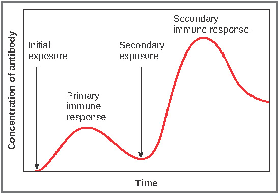
U11.1.9 Immunity depends upon the persistence of memory cells.
Immunity develops when the immune system is challenged by a specific antigen and produces antibodies and memory cells in response. Memory cells ensure that the second time an antigen is encountered, the body is ready to respond rapidly by producing more antibodies at a faster rate.
UNDERSTANDINGS:
U11.1.1 Every organism has unique molecules on the surface of its cells.
- All organisms have unique molecules or markers on the outer surface of the plasma membrane of their cells
- These highly variable molecules are generally glycoproteins and they identify a cell as being “self” or “non-self”
- These markers are called major histocompatibility complexes (MHC)
- These MHC proteins are genetically determined and are unique to that individual
U11.1.2 Pathogens can be species-specific although others can cross species barriers.
- Invading organisms such as a virus or bacterium that enters the body and causes a disease are known as pathogens
- Pathogens are generally species specific, for example, humans are the only known organisms susceptible to pathogens such as polio, syphilis, measles and gonorrhea but are resistant to many pathogens that infect other organisms
- However, there are pathogens that can cross this species barrier and infect a range of hosts, such as the Rabies virus, bird flu and the Bubonic plague
- Diseases from other animals that can infect or be transmitted to humans is called Zoonosis
- The passing of diseases from different species is a growing global health concern
U11.1.3 B lymphocytes are activated by T lymphocytes in mammals.
- When a pathogen enters the blood, the specific antigen on the surface of the membrane is identified.
- Specific phagocytes known as macrophages recognize a pathogen as a foreign entity because of the antigens on the surface.
- The macrophage engulfs and partially destroys the pathogen.
- The macrophage takes the antigens from the destroyed pathogen and displays them on the surface of the cell bound to a membrane protein called a MHC protein (called antigen presentation).
- Specific T-lymphocytes receptors recognize and bind to the antigen presented by the macrophage, thus activating the T-lymphocyte.
- The activated T-cell binds to a B-lymphocyte specific to the antigen; activating the B-cell through the binding and the release of a signaling protein
U11.1.4 Activated B cells multiply to form clones of plasma cells and memory cells.
- The active B-cells begin to clone themselves (by mitosis) producing cloned plasma B cells that produce antibodies and memory cells. The generation of large numbers of plasma cells that produce one specific antibody type is known as clonal selection. Memory cells remain in the blood in case a second infection occurs to provide long term protection and a quick response to the new infection.
- The plasma cells created produce and release mass amounts of antibodies into the bloodstream.
- These antibodies surround and bind to the antigens on the foreign pathogens.
- Through a variety of different methods the pathogens are destroyed by the antibodies and other white blood cells.
U11.1.5 Plasma cells secrete antibodies.
- As stated above, plasma cells are specialized B lymphocytes (called B cells as they develop in the bone marrow) that secrete a large amount of antibodies during a selective immune response.
- Since they are a cell that produces and secretes a large number of antibodies (proteins), they contain an extensive amount of rER, ribosomes and mitochondria (for energy).
U11.1.6 Antibodies aid the destruction of pathogens.
- Agglutination – Antibodies cause the sticking together of pathogens by attaching to the antigens on the surface. These clumped masses of pathogens are then easily ingested and destroyed by phagocytes. The large agglutinated mass can be filtered by the lymphatic system and then phagocytised.
- Opsonisation – Antibodies make pathogens recognizable by binding to them and linking them to phagocytes.
- Neutralisation of toxins – Antibodies bind to toxins produced by pathogens in the blood plasma preventing them from affecting susceptible cells.
- Complement activation – The complement system is a collection of proteins which ultimately lead to the perforation of the membranes of pathogens. Antibodies bound to the surface of a pathogen activate a complement cascade which leads to the formation of a “membrane attack complex” that forms a pore in the membrane of the pathogen allowing water and ions to enter into the cell, causing the cell to lyse.
- Neutralisation of viruses and bacteria – Antibodies can bind to the surface of viruses, preventing them from entering host cells.
U11.1.7 White cells release histamine in response to allergens.
U11.1.8 Histamines cause allergic symptoms.
Histamine is an important protein involved in many allergic reactions. Allergies are caused by an immune response to a normally innocuous substance (i.e. pollen, dust) that comes in contact with lymphocytes specific for that substance, or antigen. In many cases, the lymphocyte triggered to respond is a mast cell. For this response to occur, a free-floating IgE (an immunoglobulin associated with allergic response) molecule specific to the antigen must first be attached to cell surface receptors on mast cells. Antigen binding to the mast cell-attached IgE then triggers the mast cell to respond. This response often includes the release of histamine. It causes contraction of smooth muscle and dilation of capillaries.
Histamine causes many of the symptoms of allergies, such as a runny nose or sneezing. When a person is allergic to a particular substance, such as a food or dust, the immune system mistakenly believes that this usually harmless substance is actually harmful to the body. In an attempt to protect the body, the immune system starts a chain reaction that prompts some of the body's cells to release histamine and other chemicals into the bloodstream. The histamine then acts on a person's eyes, nose, throat, lungs, skin, or gastrointestinal tract, causing allergy symptoms.
NOTE: this is why people with allergies take antihistamines!
U11.1.9 Immunity depends upon the persistence of memory cells.
Immunity develops when the immune system is challenged by a specific antigen and produces antibodies and memory cells in response. Memory cells ensure that the second time an antigen is encountered, the body is ready to respond rapidly by producing more antibodies at a faster rate.
U11.1.10 Vaccines contain antigens that trigger immunity but do not cause the disease.
- Vaccines are introduced to the body usually through an injection but can be administered through orally or through a nasal spray
- Vaccines contain a live attenuated (weakened) or killed version of the pathogen, itstoxins or one of its surface antigens.
- Vaccines stimulate a primary immune response
- If the body encounters the actually pathogen, it will be destroyed right away by theantibodies during a secondary immune response
- Vaccines has made great contributions towards public health through the prevention of many deadly or dangerous diseases such as tuberculosis, measles and smallpox
U11.1.11 Fusion of a tumour cell with an antibody-producing plasma cell creates a hybridoma cell.
Monoclonal antibodies are highly specific, purified antibodies that are produced by a clone of cells, derived from a single cell. They recognise only one antigen. To produce the clone of cells that will manufacture a monoclonal antibody, the antigen recognised by the antibody is injected into a mouse, or other mammal. In response to this challenge, the mouse's immune system makes plasma B cells that are capable of producing the desired antibody. Plasma cells are removed from the spleen of the mouse. They will be of many different types with only some producing the desired antibody.
The B cells are fused with cancer cells called myeloma cells. The cells formed by fusion of plasma B cells and myeloma cells are called hybridoma cells.
U11.1.12 Monoclonal antibodies are produced by hybridoma cells.
- Monoclonal antibodies are identical antibodies produced by clones of a single parent immune cell that are specific to one type of antigen.
- A laboratory animal such as a mouse is injected with a specific antigen thatcorresponds with the needed antibodies.
- After the animal goes through a primary immune response, a plasma B-cell cell that produces the required antibody is removed from the spleen.
- Myeloma (cancer) cells are cultured in a petri dish.
- These dividing myeloma cells are mixed together with the plasma B-cells and are treated to promote a fusion between the two cells, forming a cell called a hybridoma.
- The successful hybridomas have characteristics of both cells; produce antibodies and divide rapidly for a long time.
- These hybridoma cells are cultured and allowed to divide, producing many clone cellsthat are able to produce large amounts of antibodies.
- Monoclonal antibodies can be extracted and used for many different applications.
APPLICATION:
A11.1.1 Smallpox was the first infectious disease of humans to have been eradicated by vaccination.
A11.1.2 Monoclonal antibodies to HCG are used in pregnancy test kits.
A11.1.3 Antigens on the surface of red blood cells stimulate antibody production in a person with a different blood group.
SKILL:
S11.1.1 Analysis of epidemiological data related to vaccination programmes.
Essential idea: The roles of the musculoskeletal system are movement, support and protection.
11.2 Movement
UNDERSTANDINGS:
U11.2.1 Bones and exoskeletons provide anchorage for muscles and act as levers.
U11.2.2 Synovial joints allow certain movements but not others.
U11.2.3 Movement of the body requires muscles to work in antagonistic pairs.
Skeletal muscles occurs in pairs that are antagonistic. This means that when one contracts, the other relaxes. Antagonistic muscles produce opposite movements at a joint. For example, in the elbow, the triceps extends the forearm while the biceps flex the forearm.
U11.2.4 Skeletal muscle fibres are multinucleate and contain specialized endoplasmic reticulum.
U11.2.5 Muscle fibres contain many myofibrils.
Within each muscle fibre there are many parallel, elongated structures called myofibrils. These have alternating light and dark bands, which give striated muscle its strips. In the centre of each light band is a disc-shaped structure, referred to as the Z-line.
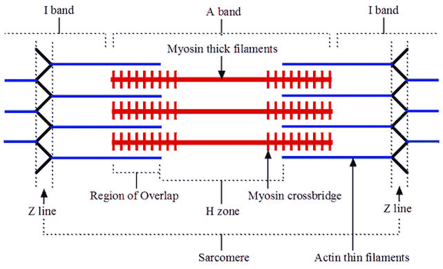
U11.2.6 Each myofibril is made up of contractile sarcomeres.
Myofibrils – rod-shaped parallel bodies consisting of actin and myosin filaments
U11.2.7 The contraction of the skeletal muscle is achieved by the sliding of actin and myosin filaments.
U11.2.8 ATP hydrolysis and cross bridge formation are necessary for the filaments to slide.
U11.2.9 Calcium ions and the proteins tropomyosin and troponin control muscle contractions.
UNDERSTANDINGS:
U11.2.1 Bones and exoskeletons provide anchorage for muscles and act as levers.
- Bones act as levers so the body can move and provide structural support (skeleton).
- Ligaments are strong bands that connect bone to bone strengthening the joint during movement.
- Tendons have dense connective tissue that connects muscles to bones, allowing movement of the bone when a muscle contracts.
- Muscles provide the force for movement by contracting (shortens the muscle fibers)
- The joint acts as a pivot point or a fulcrum
- The force applied (when the muscle contracts) is called the effort
- The force or load needed to overcome for movement to take place is called the resistance
- Levers are classified by first, second, and third class, depending upon the positions among the fulcrum, the effort, and the resistance.
- First-class levers have the fulcrum in the middle, like a seesaw. An example of a first class lever is when a human nods their head (top of the spinal column is the fulcrum, the effort force is provided by the muscles in the back of the neck, and the resistance is weight of the head).
- Second-class levers have a resistance in the middle, like a load in a wheel-barrow. The body acts as second class lever when engaged in pushup or calf raise. During a calf raise ball of foot is fulcrum, the body’s mass is the resistance and the effort is applied by calf muscle.
- Third-class levers have the effort from the muscle in the middle of the lever. The majority of the human body's musculoskeletal levers are third class. These levers are built for speed and range of motion. Muscle attachments are usually close to the fulcrum. In the example of the arm, the effort force is provided by the contraction of the biceps, the fulcrum is the elbow joint and the resistance would be provided by whatever weight is being lifted.
- Exoskeletons in insects and crustaceans can facilitate the movement by providing an anchorage for muscles; similarly to how bones provide anchorage for animals with internal skeletons.
U11.2.2 Synovial joints allow certain movements but not others.
- The type of joint determines the amount of movement that is possible
- For ball and socket joints, such as the hip or the shoulder, movement through all three planes are possible. At the hip joint, the head of the femur is the ball the fits into the socket of the pelvis. The movements possible at the joint are flexion, extension, rotation, abduction and adduction.
- For hinge joints, such as the knee, flexions (bending) and extensions (straightening) are the possible movements (movement in one plane); however, slight side to side movements are possible.
U11.2.3 Movement of the body requires muscles to work in antagonistic pairs.
Skeletal muscles occurs in pairs that are antagonistic. This means that when one contracts, the other relaxes. Antagonistic muscles produce opposite movements at a joint. For example, in the elbow, the triceps extends the forearm while the biceps flex the forearm.
U11.2.4 Skeletal muscle fibres are multinucleate and contain specialized endoplasmic reticulum.
- Skeletal muscles are composed of bundles of muscle fibers and have a striped appearance because of areas of thick and thin filaments (myosin and actin)
- Muscle cells have many nuclei and are long because the embryonic muscle cells fuse together
- Muscle fibers are composed of many parallel elongated fibers called myofibrils
- A modified endoplasmic reticulum, called the sarcoplasmic reticulum (fluid-filled membranous sacs), extends throughout the muscle fibre, wrapping around each myofibril, sending a signal to the all parts of the muscle fibre to contract at the same time
- The sarcoplasmic reticulum stores calcium - between myofibrils are large numbers of mitochondria, which provide ATP needed for contractions.
U11.2.5 Muscle fibres contain many myofibrils.
Within each muscle fibre there are many parallel, elongated structures called myofibrils. These have alternating light and dark bands, which give striated muscle its strips. In the centre of each light band is a disc-shaped structure, referred to as the Z-line.
U11.2.6 Each myofibril is made up of contractile sarcomeres.
Myofibrils – rod-shaped parallel bodies consisting of actin and myosin filaments
- Sarcolemma – plasma membrane of the muscle cell
- Mitochondria – large numbers; found dispersed around individual myofibrils
- Lies between two Z-lines which are dense protein discs
- Contains the thick filament (myosin) and thin filament (actin)
- Myosin contains a head which binds to the binding site on the actin; interaction between myosin and actin (cross-bridge) is responsible for muscle contraction
- Myosin is seen as dark bands while actin is seen as light bands
- Actin filaments are attached to a Z-line at one end
- Myosin filaments are interdigitated with actin filaments at both ends and occupy the centre of the sarcomere
- Each mysosin filament is surrounded by six actin filaments
- A bands contain a full length of myosin and some of the actin filaments
- I bands contain only actin filaments
U11.2.7 The contraction of the skeletal muscle is achieved by the sliding of actin and myosin filaments.
U11.2.8 ATP hydrolysis and cross bridge formation are necessary for the filaments to slide.
U11.2.9 Calcium ions and the proteins tropomyosin and troponin control muscle contractions.
- the action potential triggers the opening of calcium-gated ion channels
- calcium ions floods into the presynaptic terminal
- this causes vesicles of neurotransmitters to migrate down to the presynaptic membrane and they fuse with the membrane
- releases neurotransmitters into the cleft
- the neurotransmitters bind to the receptors on the muscle
- depolarises the myofibril membrane
- the calcium ions are released from the sarcoplasmic reticulum
- calcium ions bind to the troponin and displaces tropomyosin
- myosin head can then bind to the binding site on actin
- because the actin filaments are anchored to the Z-lines, the sarcomere shortens from both sides when actin filaments slide along myosin filaments
- the sliding action = the myosin actually pulls the actin along its length
- the cross bridges of the myosin filaments attach to the actin filaments and exert force on them to move - this action is known as the sliding filament mechanism of muscle contraction
- the sarcomeres shorten without the thick or thin filaments changing in length
- a contraction begins when a bound ATP molecule is hydrolysed to ADP + inorganic phosphate
- this causes the myosin head to extend and can attach to a binding site on actin forming a cross bridge
- an action called the power stroke is triggered, allowing myosin to pull the actin filament toward to M-line, thereby shortening the sarcomere
- ADP and inorganic phosphate are released during the power stroke
- the myosin remains attached to actin until a new molecule of ATP binds, freeing the myosin to either go through another cycle of binding and more contraction, or remain unattached to allow the muscle to relax
- muscle contractions are controlled by the action of calcium
- the thin actin filaments are associated with regulatory proteins called troponin and tropomyosin
- when a muscle is relaxed, tropomyosin blocks the cross bridge binding sites on the actin
- when calcium ion levels are high enough and ATP is present, calcium ions bind to the troponin which displaces tropomyosin, exposing the myosin binding sites on actin
- this allows myosin to attach to a binding site on actin, forming a cross bridge
- calcium ions are stored in the sarcoplasmic reticulum and are released in response to signals from the nervous system to contract
- neurotransmitter molecules are released from a neuron and bind to receptors which depolarises the membrane of the muscle fibre
- the electrical impulse travels down the T-tubules and opens calcium stores
- calcium ions flow to the myofibrils where they trigger a muscle contraction
- as the actin and myosin slide along each other, the entire sarcomere shortens, as the Z-lines draw closer to the M-line
- as the sarcomeres in myofibrils contract, the entire muscle fibre will shorten.
APPLICATION:
A11.2.1 Antagonistic pairs of muscles in an insect leg.
SKILL:
S11.2.1 Annotation of a diagram of the human elbow.
S11.2.2 Drawing labelled diagrams of the structure of a sarcomere.
S11.2.3 Analysis of electron micrographs to find the state of contraction of muscle fibres.
NOTE: the A band in the diagram below = the dark band and the I band = the light band
Essential idea: All animals excrete nitrogenous waste products and some animals also balance water and solute concentrations.
11.3 The kidney and osmoregulation
UNDERSTANDINGS:
U11.3.1 Animals are either osmoregulators or osmoconformers.
- osmolarity refers to the solute concentration of a solution
- many animals are known as osmoregulators because they maintain constant internal solute concentration, even when living in marine environments with very different osmolarities
- all terrestrial animals, freshwater animals and some marine organisms like bony fish are osmoregulators
- typically these organisms maintain their solute concentration at about one third of the concentration of seawater and about 10 times that of fresh water
- osmoconformers are animals whose internal solute concentration tends to be the same as the concentration of solutes of the environment
U11.3.2 The Malpighian tubule system in insects and the kidney carry out osmoregulation and removal of nitrogenous wastes.
- arthropods have circulating fluid, known as hemolymph, that combines the characteristics of tissue fluid and blood
- osmoregulation is a form of homeostasis whereby the concentration of hemolymph, or blood in the case of animals with closed circulatory systems, is kept within a certain range
- when animals break down amino acids, the nitrogenous waste product is toxic and needs to be excreted
- in insects, the waste product is usually in the form of uric acid and in mammals it is in the form of urea
- insects have tubes that branch off from their intestinal tract
- these are known as Malpighian tubules
- cells lining the tubules actively transport ions and uric acid from the hemolymph into the lumen of the tubules
- this draws water by osmosis from the hemolymph through the walls of the tubules into the lumen
- the tubules empty their contents into the gut
- in the hindgut most of the water and salts are reabsorbed while the nitrogenous waste is excreted with the feces
U11.3.3 The composition of blood in the renal artery is different from that in the renal vein.
- renal artery = blood enters the kidney
- renal vein = blood leaves the kidney
Substances that are present in HIGHER amounts in the renal artery than the renal vein include:
- toxins and other substances that are ingested and absorbed but are not fully metabolised by the body e.g. betain pigments in beets and also drugs
- excretory waste products including nitrogenous waste products, mainly urea
Other things removed from the blood by the kidney that are not excretory products include:
- excess water, produced by cell respiration or absorbed from food in the gut
- excess salt, absorbed from food in the gut
NOTE: the kidney is the filter of the body - thus the substances that need to be removed from the body are present in higher amounts in the blood vessel entering the kidney (the renal artery).
- the metabolic activity of the kidney itself also causes a difference between the composition of blood in the renal artery and the renal vein
- blood leaving the kidney through the renal vein is deoxygenated relative to the renal artery because kidney metabolism requires oxygen
- it also has a higher partial pressure of CO2 because this is a waste product of metabolism
- even though glucose is normally filtered and then entirely reabsorbed, some glucose is used by the metabolism of the kidney and therefore the concentration is slightly lower in the renal vein compared to the renal artery
U11.3.4 The ultrastructure of the glomerulus and Bowman’s capsule facilitate ultrafiltration.
- blood in capillaries is at high pressure in many of the tissues of the body, and the pressure forces some of the plasma out through the capillary wall, to form tissue fluid
- in the glomerulus of the kidney, the pressure in the capillaries is particularly high and the capillary wall is particularly permeable, so the volume forced out is about 100 times greater than in other tissues
- the fluid forced out is called glomerular filtrate
- the concentration of Na+ ions, Cl- ions, glucose and urea all remain about the same in both the composition of blood plasma and the filtrate, however protein (mg) differs enormously
- this separation of particles differing in size by a few nanometers is called ultrafiltration
- all particles with a relative molecular mass below 65,000 atomic mass units pass through
- the permeability to larger molecules depends on their shape and charge
- almost all proteins are retained in the blood, along with all blood cells
The glomerulus:
- the basement membrane separates the capillaries
- there are gaps in the wall of the capillaries called fenestrations
- smaller projections from the membrane are podocyte food processes, which attach the podocytes (specialised epithelial cells) to the membrane
- the podocytes function as a barrier through which waste products are filtered from the blood
There are three main parts to the ultrafiltration system:
1. Fenestrations between the cells in the wall of the capillaries - these are about 100nm in diameter and they allow fluid to escape, but not blood cells
2. The basement membrane that covers and supports the wall of the capillaries - it is made of negatively-charged glycoproteins, which form a mesh - it prevents plasma proteins from being filtered out, due to their size and negative charges
3. Podocytes forming the inner wall of the Bowman's capsule - these cells have extensions that wrap around the capillaries of the glomerulus and many short side branches called foot processes - very narrow gaps between the foot processes help prevent small molecules from being filtered out of the blood in the glomerulus
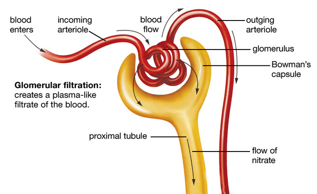
NOTE: the incoming arteriole in the diagram above is called the afferent arteriole and the outgoing arteriole is called the efferent arteriole
U11.3.5 The proximal convoluted tubule selectively reabsorbs useful substances by active transport.
- the glomerular filtrate flows into the proximal convoluted tubule
- the volume of glomerular filtrate produced per day is huge - about 180dm-3
- most reabsorption of glucose and other substances mainly occurs in the first part of the nephron - the proximal convoluted tubule
Methods use to reabsorb substances in the proximal convoluted tubule include:
Sodium ions:
- moved by active transport from filtrate to space outside the tubule
- they then pass to the peritubular capillaries
- pump proteins are located in outer membrane of tubule cells
Chloride ions:
- attracted from filtrate to space outside the tubule because of charge gradient set up by active transport of sodium ions
Glucose:
- co-transported out of filtrate and into fluid outside the tubule, by co-transporter proteins in outer membranes of tubules cells
- sodium ions move down concentration gradient from outside tubule into tubule cells
- this provides energy for glucose to move at the same time to fluid outside the tubule
- the same process is used to reabsorb amino acids
By the end of the proximal tubule all glucose and amino acids and 80% of the water, sodium and other mineral ions have been absorbed.
U11.3.6 The loop of Henle maintains hypertonic conditions in the medulla.
-
U11.3.7 ADH controls reabsorption of water in the collecting duct.
U11.3.8 The length of the loop of Henle is positively correlated with the need for water conservation in animals.
U11.3.9 The type of nitrogenous waste in animals is correlated with evolutionary history and habitat.
Essential idea: Sexual reproduction involves the development and fusion of haploid gametes.
11.4 Sexual reproduction
UNDERSTANDINGS:
U11.4.1 Spermatogenesis and oogenesis both involve mitosis, cell growth, two divisions of meiosis and differentiation.
OOGENESIS:
- oogenesis is the production of egg cells in the ovaries
- oogenesis starts in the ovaries of a female fetus
- germ cells in the fetal ovary divide by mitosis and the cells formed move to distribute themselves through the cortex of the ovary
- when the fetus is four/five months old, these cells grow and start to divide by meiosis
- by the seventh month, they are still in the first division of meiosis and a single layer of cells, called follicle cells, has formed around them
- no further development takes place until after puberty
- the cell that has started to divide by meiosis, together with the surrounding follicle cells, is called a primary follicle
- there are about 400,000 in the ovaries at birth
- no more primary follicles are produced, but at the start of eaach menstrual cycle, a small batch are stimulated to develop by FSH
- usually one goes on to become a mature follicle, containing a secondary oocyte
SPERMATOGENESIS:
- spermatogenesis is the production of sperm
- it happens in the testes, which are composed of a mass of narrow tubes, called seminiferous tubules, with small groups of cells filling the gaps between the tubules
- these gaps are called interstices, so the cells in them are interstital cells
- they are sometimes called Leydig cells
- the seminiferous tubules are also made of cells
- the outer layer of cells is called the germinal epithelium, with the most mature stages closest to the fluid-filled centre of the seminiferous tubule
- cells that have developed tails are called spermatoza
- in the wall of the tubule are large nurse cells, called Sertoli cells, which nourish the sperm cells
U11.4.2 Processes in spermatogenesis and oogenesis result in different numbers of gametes with different amounts of cytoplasm.
U11.4.3 Fertilization in animals can be internal or external.
U11.4.4 Fertilization involves mechanisms that prevent polyspermy.
U11.4.5 Implantation of the blastocyst in the endometrium is essential for the continuation of pregnancy.
U11.4.6 HCG stimulates the ovary to secrete progesterone during early pregnancy.
U11.4.7 The placenta facilitates the exchange of materials between the mother and fetus.
U11.4.8 Estrogen and progesterone are secreted by the placenta once it has formed.
U11.4.9 Birth is mediated by positive feedback involving estrogen and oxytocin.