essential idea: the evolution of multicellular organisms allowed cell SPECIALISATION and cell replacement
1.1 Introduction to cells
UNDERSTANDINGS:
U1.1.1 According to the cell theory, living organisms are composed of cells.
Cell theory states: living things are composed of one or more cells; the cell is the basic unit of life; cells arise from existing cells.
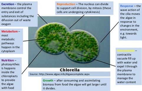
U1.1.2 Organisms consisting of only one cell carry out all functions of life in that cell.
Unicellular organisms (such as amoeba, paramecium, euglena and bacterium) are the smallest organisms capable of independent life.
All living things share 7 basic characteristics:
Movement: Living things show movement, either externally or internally
Reproduction: Living things produce offspring, either sexually or asexually
Sensitivity: Living things can respond to and interact with the environment
Growth: Living things can change size / shape
Respiration: Living things use substances from the environment to create energy
Excretion: Living things exhibit the removal of wastes
Nutrition: Living things exchange materials and gases with the environment
U1.1.3 Surface area to volume ratio is important in the limitation of cell size.
SA:V ratio effectively limits the size of cells. In the cell, the rate of heat and waste production and rate of resource consumption are functions that depend on its volume. Most of the chemical reactions occur in the interior of the cell and its size affects the rate of these reactions. The surface of the cell, the membrane, controls what materials move in and out of the cell. Cells with more SA per unit volume are able to move more materials in and out of the cell, for each unit of volume of the cell.
As the width of an object such as a cell increases, the SA also increase but at a much slower rate than the volume. This means that a large cell has relatively less SA to bring in needed materials and to rid the cell of waste than a small cell. Because of this, cells are limited as to the size they can attain and still be able to carry out the functions of life. Thus, large animals do not have large/r cells, they have more cells. Cells that are larger in size have modifications that allow them to function efficiently. This is accomplished by shape changes such as from spherical to long and thin. Some cells have infoldings or outfoldings to increase their surface relative to their volume.
Summary:
- Substances need to be taken in to the cell to fuel reactions & waste products need to be removed
- Increase in cell size leads to increase in chemical reactions --> more substances needed in and more substances needing to be removed
- Surface area affects the rate at which particles enter and exit the cell
- Volume affects the rate of the chemical activities
- When the volume increases so does the surface area but not to the same extent
- As the cell gets larger, its surface area to volume ratio gets smaller
- If the ratio gets too small, particles will not be able to enter and exit the cell fast enough
- Results in accumulation of waste products and overheating of the cell
U1.1.4 Multicellular organisms have properties that emerge from the interaction of their cellular components.
One of the most intensively studied researched multicellular organisms is a worm called Caenorhabditis elegans. It has no common name and lives unseen in decomposing organic matter - it is one mm in length and composed of 959 cells. It has a mouth, pharynx, intestine and anus. It is a hermaphrodite so it has both male and female reproductive organs. Almost a third of the cells are neurons, or nerve cells. Most of these neurons are located at the front end of the worm in a structure that can be regarded as the animal's brain.
Although the brain in C. elegans coordinates responses to the worm's environment, it does not control how individual cells develop. The cells in this and other multicellular organisms can be regarded as cooperative groups, without any cells in the group acting as a leader or supervisor. The characteristics of the whole organism, including the fact that it is alive, are known as emergent properties.
Emergent properties arise from the interaction of the component parts of a complex structure i.e. the whole is greater than the sum of its parts.
Multicellular organisms are capable of completing functions that individual cells could not undertake - this is due to the interaction between cells producing new functions.
In multicellular organisms:
- Cells may group together to form tissues
- Organs are then formed from the functional grouping of multiple tissues
- Organs that interact may form organ systems capable of carrying out specific body functions
- Organ systems carry out the life functions required by an organism.
U1.1.5 Specialized tissues can develop by cell differentiation in multicellular organisms.
In multicellular organisms different cells perform different functions. This is sometimes called division of labour. For example, the job of red blood cells is to carry oxygen, and the function of a rod cell in the retina of the eye is to absorb light and then transmit impulses to the brain. Often a group of cells specialise in the same way to perform the same function - called a tissue.
By becoming specialised, the cells in a tissue can carry out their role more effectively than if they had many different roles. They can develop the ideal structure, with the enzymes needed to carry out all of the chemical reactions associated with the function. The development of cells in different ways to carry out specific functions is called differentiation. In humans, 220 distinctively different highly specialised cell types have been recognised, all of which develop by differentiation.
U1.1.6 Differentiation involves the expression of some genes and not others in a cell’s genome.
Every cell in a multicellular organisms contains all the genes of that organism. However, the genes that are activated vary from cell to cell. The reason we have different types of cells in our body (the cells in your eyes are not the same as the ones that make up your hair) is because different genes are activated in different cells. For example, the gene that produces keratin will be active in hair and nail cells. Keratin is the protein which makes up hair and nails. Genes encode for proteins and the proteins affect the cell’s structure and function so that the cell can specialize. This means cells develop in different ways. This is called differentiation. Differentiation depends on gene expression which is regulated mostly during transcription. It is an advantage for multicellular organisms as cells can differentiate to be more efficient unlike unicellular organisms who have to carry out all of the functions within that one cell.
Summary:
- All cells of an individual organisms share an identical genome - each cell contains the entire set of genetic instructions for that organism
- The activation of different instructions (genes) within a given cell by chemical signals will cause it to differentiate from other cells like it
- Differentiation is the process during development whereby newly formed cells become more specialised and distinct from one another as they mature
- Active genes are usually packaged in an expanded and accessible form (euchromatin), while inactive genes are mainly packaged in a condensed form (heterochromatin)
- Differentiated cells will have different regions of DNA packaged as heterochromatin and euchromatin depending on their function.
U1.1.7 The capacity of stem cells to divide and differentiate along different pathways is necessary in embryonic development and also makes stem cells suitable for therapeutic uses.
Stem cells are cells that are not fully differentiated but have the ability to divide and differentiate into different types of cells (e.g. one stem cell can differentiate into a blood cell, a liver cell or a kidney cell). Stem cells are necessary in embryonic development as all the cells in the adult organism stem from the embryonic stem cells (hence the name).
When an egg cell and a sperm cell fuse they form a zygote. This zygote then starts to divide to form a two celled embryo, then a 4 celled embryo, then an 8 celled embryo... and so on. Early in the early stages of the embryo the cells are stem cells because they are able to become any type of cell within the organism (therefore they are said to be totipotent stem cells --> arises from the word total) and can divide multiple times.
Gradually as the embryo cells divide they start to become committed to a specific path so for example some cells become committed to being kidney cells. So even though they retain their ability to differentiate, they can only differentiate into kidney cells (and no longer liver or blood cells for example), therefore they are no longer stem cells and are said to be unipotent (uni = one). However a small amount of stem cells are still found in the adult organism, e.g. stem cells can still be found in the skin, bone marrow and liver.
Even though these stem cells are not able to differentiate into all the different types of cells in the organism (unlike the embryo stem cells that are totipotent these are only pluripotent --> arises from the word plural --> many but not total) they are still able to differentiate into a range of different cells so in the case of stem cells within the bone marrow, these are not able to differentiate into liver cells for example but are able to differentiate into a multitude of white blood cells and into red blood cells. So even though these remaining stem cells are not as effective as embryonic stem cells they still greatly benefit the adult organism in terms of regeneration and repair of damaged tissue.
Since stem cells are able to differentiate multiple times and into different cell types, they are ideal for therapeutic use in degenerative diseases or tissue repair.
Examples of therapeutic uses of stem cells:
1. Retinal cells: Replace dead cells in retina to cure diseases like glaucoma and macular degeneration.
2. Skin cells: Graft new skin cells to replace damaged cells in severe burn victims.
3. Nerve cells: Repair damage caused by spinal injuries to enable paralysed victims to regain movement.
4. Blood cells: Bone marrow transplants for cancer patients who are immuno-compromised as a result of chemotherapy.
APPLICATION:
A1.1.1 Questioning cell theory using atypical examples, including striated muscle, giant algae and aseptate fungal hyphae.
Striated muscle:
- Challenges the idea that a cell has one nucleus
- Muscle cells have more than one nucleus per cell
- Muscle cells called fibres can be very long
- They are surrounded by a single plasma membrane but they are multi-nucleated
- Does not conform to the standard view of a small, single nucleus within a cell
Giant algae:
- Acetabularia is a single-celled organism that challenges both the idea that a cell must be simple in structure and small in size
- Gigantic in size (5-100 mm)
- Complex in form - consists of 3 anatomical parts i.e. bottom rhizoid, long stalk, top umbrella of branches
- Single nucleus is located in the rhizoid
Aseptate fungal hyphae:
- Challenges the idea that a cell is a single unit
- Fungal hyphae are very large with many nuclei and a continuous cytoplasm
- The tubular system of hyphae form dense networks called mycelium
- Cell walls are composed of chitin
- No end cell wall or membrane
A1.1.2 Investigation of functions of life in Paramecium and one named photosynthetic unicellular organism.
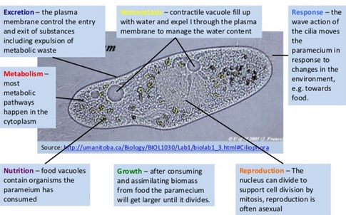
NOTE: the yellow writing says "Homeostasis" - maintaining a constant internal environment.
Source: http://fr.slideshare.net/diverzippy/biok-notes-11-introduction-to-cells
A1.1.3 Use of stem cells to treat Stargardt's disease and one other named condition.
Stargardt's disease:
What is it?
Stargardt's disease — also called fundus flavimaculatus or Stargardt's macular dystrophy (SMD) — affects approximately one in 10,000 people and is characterized by central vision loss early in life. (Some researchers believe a distinction should be made between Stargardt's disease and fundus flavimaculatus, because they say each describes a different variant of the eye disease.)
Stargardt's generally refers to a group of inherited diseases causing light-sensitive cells in the inner back of the eye (retina) to deteriorate, particularly in the area of the macula where fine focusing occurs. Central vision loss also occurs, while peripheral vision usually is retained.
Stargardt's disease is diagnosed by the presence of small, yellowish spots of deteriorating tissue (drusen) sloughed off from the colored or outer covering of the retina (retinal pigment epithelium). Progressive vision loss eventually leads to blindness in most cases.
What is the treatment?
Embryonic stem cells are unique in that they have the potential to develop (differentiate) into any type of adult body cell. Most embryonic stem cells are taken from embryos that have been fertilised in vitro in the laboratory (IVF), and the cells are then cultured in the laboratory, remaining in their undifferentiated form unless researchers put them under conditions that will allow them to change into different cell types.
Under laboratory conditions it has been possible to develop retinal cells from these cells, which is a significant advance and could potentially be translated into new treatments for patients who have otherwise untreatable visual loss due to damage to their own retinal cells.
During an operation lasting up to an hour, the stem-cell-derived retinal cells will be transplanted into adults with severe sight impairment as a result of Stargardt’s disease, but who are otherwise healthy.
Other conditions:
A bone marrow transplant, also known as a haemopoietic stem cell transplant, replaces damaged bone marrow with healthy bone marrow stem cells.
Bone marrow is a spongy tissue found in the hollow centres of some bones. It contains specialist stem cells, which produce the body's blood cells.
Stem cells in bone marrow produce three important types of blood cells:
Why are bone marrow transplants needed?
Bone marrow transplants are often needed to treat conditions that damage bone marrow. If bone marrow is damaged, it is no longer able to produce normal blood cells. The new stem cells take over blood cell production.
Conditions that bone marrow transplants are used to treat include:
A bone marrow transplant has five stages. These are:
Risks?
Bone marrow transplants are complicated procedures with significant risks.
In some cases, the transplanted cells (graft cells) recognise the recipient's cells as "foreign" and try to attack them. This is known as graft versus host disease (GvHD).
The risk of infection is also increased because your immune system is weakened when you're conditioned (prepared) for the transplant.
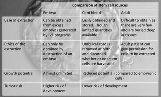
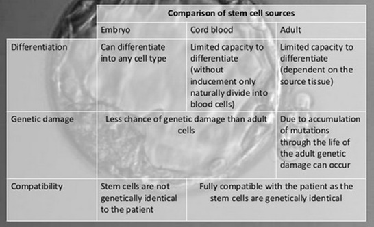
A1.1.4 Ethics of the therapeutic use of stem cells from specially created embryos, from the umbilical cord blood of a new-born baby and from an adult’s own tissues.
Stargardt's disease:
What is it?
Stargardt's disease — also called fundus flavimaculatus or Stargardt's macular dystrophy (SMD) — affects approximately one in 10,000 people and is characterized by central vision loss early in life. (Some researchers believe a distinction should be made between Stargardt's disease and fundus flavimaculatus, because they say each describes a different variant of the eye disease.)
Stargardt's generally refers to a group of inherited diseases causing light-sensitive cells in the inner back of the eye (retina) to deteriorate, particularly in the area of the macula where fine focusing occurs. Central vision loss also occurs, while peripheral vision usually is retained.
Stargardt's disease is diagnosed by the presence of small, yellowish spots of deteriorating tissue (drusen) sloughed off from the colored or outer covering of the retina (retinal pigment epithelium). Progressive vision loss eventually leads to blindness in most cases.
What is the treatment?
Embryonic stem cells are unique in that they have the potential to develop (differentiate) into any type of adult body cell. Most embryonic stem cells are taken from embryos that have been fertilised in vitro in the laboratory (IVF), and the cells are then cultured in the laboratory, remaining in their undifferentiated form unless researchers put them under conditions that will allow them to change into different cell types.
Under laboratory conditions it has been possible to develop retinal cells from these cells, which is a significant advance and could potentially be translated into new treatments for patients who have otherwise untreatable visual loss due to damage to their own retinal cells.
During an operation lasting up to an hour, the stem-cell-derived retinal cells will be transplanted into adults with severe sight impairment as a result of Stargardt’s disease, but who are otherwise healthy.
Other conditions:
A bone marrow transplant, also known as a haemopoietic stem cell transplant, replaces damaged bone marrow with healthy bone marrow stem cells.
Bone marrow is a spongy tissue found in the hollow centres of some bones. It contains specialist stem cells, which produce the body's blood cells.
Stem cells in bone marrow produce three important types of blood cells:
- red blood cells – which carry oxygen around the body
- white blood cells – which help fight infection
- platelets – which help stop bleeding
Why are bone marrow transplants needed?
Bone marrow transplants are often needed to treat conditions that damage bone marrow. If bone marrow is damaged, it is no longer able to produce normal blood cells. The new stem cells take over blood cell production.
Conditions that bone marrow transplants are used to treat include:
- severe aplastic anaemia (bone marrow failure)
- leukaemia – cancer of the white blood cells
- non-Hodgkin lymphoma – cancer of the lymphatic system
- certain genetic blood and immune system disorders – such as sickle cell anaemia, thalassaemia and some severe immune system conditions
A bone marrow transplant has five stages. These are:
- physical examination – to assess your general level of health
- harvesting – the process of obtaining the stem cells to be used in the transplant
- conditioning – preparing your body for the transplant
- transplanting the stem cells
- recovery period –during which you'll be monitored for any complications or side effects
Risks?
Bone marrow transplants are complicated procedures with significant risks.
In some cases, the transplanted cells (graft cells) recognise the recipient's cells as "foreign" and try to attack them. This is known as graft versus host disease (GvHD).
The risk of infection is also increased because your immune system is weakened when you're conditioned (prepared) for the transplant.
A1.1.4 Ethics of the therapeutic use of stem cells from specially created embryos, from the umbilical cord blood of a new-born baby and from an adult’s own tissues.
Source: http://fr.slideshare.net/diverzippy/biok-notes-11-introduction-to-cells
SKILL:
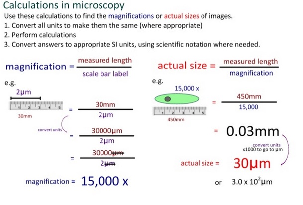
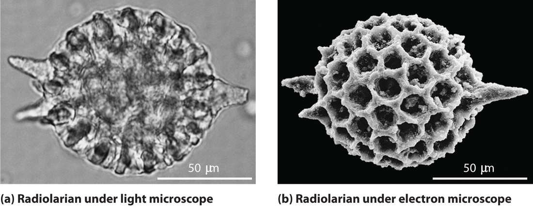
S1.1.1 Use of a light microscope to investigate the structure of cells and tissues, with drawing of cells. Calculation of the magnification of drawings and the actual size of structures and ultrastructures shown in drawings or micrographs.
Source: http://i-biology.net/ibdpbio/02-cells/cell-theory/
S1.1.1 Use of a light microscope to investigate the structure of cells and tissues, with drawing of cells. Calculation of the magnification of drawings and the actual size of structures and ultrastructures shown in drawings or micrographs.
Source: http://i-biology.net/ibdpbio/02-cells/cell-theory/
Essential idea: Eukaryotes have a much more complex cell structure than prokaryotes.
1.2 Ultrastructure of cells
UNDERSTANDINGS:
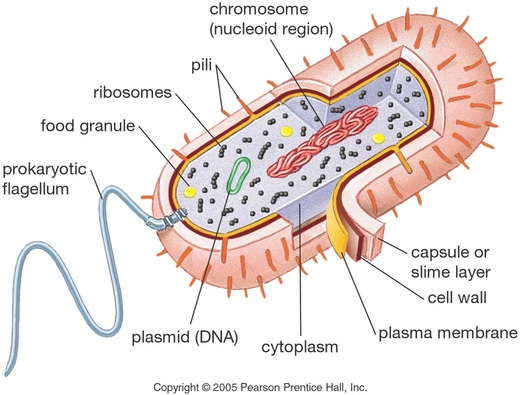
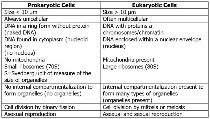
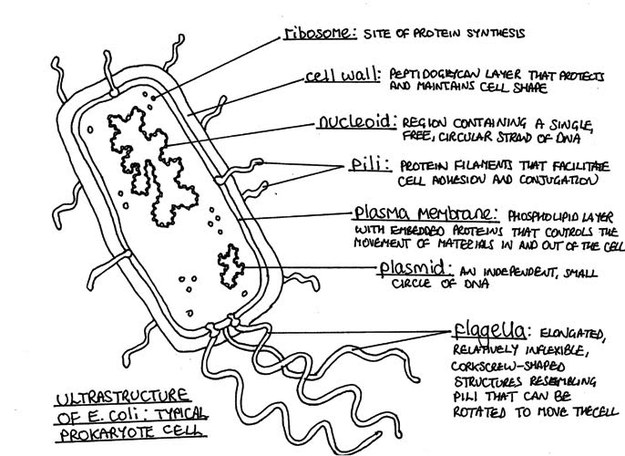
U1.2.1 Prokaryotes have a simple cell structure without compartmentalisation.
A1.2.2 Prokaryotes divide by binary fission.
Internal structures of prokaryotic cells
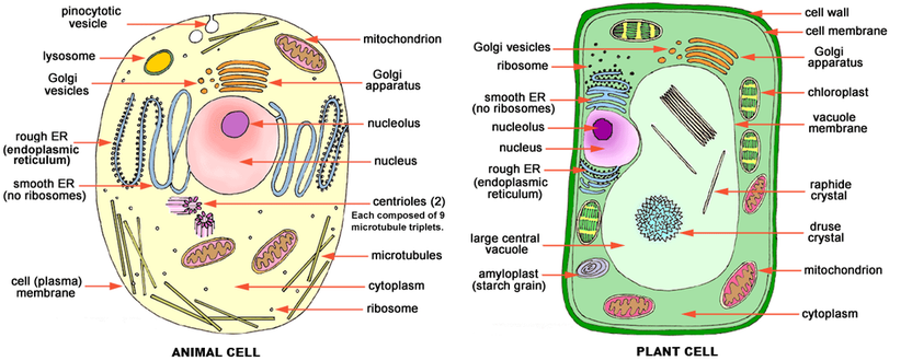
U1.2.2 Eukaryotes have a compartmentalized cell structure.
UNDERSTANDINGS:
U1.2.1 Prokaryotes have a simple cell structure without compartmentalisation.
A1.2.2 Prokaryotes divide by binary fission.
Internal structures of prokaryotic cells
- Cell wall: protects and maintains the shape of the cell - it is composed of a carbohydrate-protein complex called peptidoglycan.
- Plasma Membrane: The plasma membrane is a double-layer of phospholipids with associated proteins and other molecules. It is essentially the “bag” that holds all of the intracellular material and regulates the movement of materials into and out of the cell.
- Cytoplasm: this is the gel-like fluid that the cell is filled with, inside the plasma membrane - liquid with all of the cellular organelles suspended within. There is no compartmentalisation in the cytoplasm so all cellular processes take place in the cytoplasm.
- Cytoskeleton: it's only recently been discovered that rod-shaped bacteria and Archaea possess cytoskeletal proteins that function in a similar way to the cytoskeleton of eukaryotic cells. This scaffolding provides structural support to the cell and plays a role in cell-division.
- Ribosomes (70S): all cells, both prokaryotic and eukaryotic, have multiple ribosomes within. Ribosomes are the tiny protein-making machines of the cell.
- Pili and flagella: Pili's funtion is to join bacterial cells in preparation for the transfer of DNA from one cell to another (sexual reproduction). Flagella are longer than pili and allow the cell motility.
- Nucleoid region: this region is non-compartmentalised and contains a single, long, continuous, circular thread of DNA. In addition to the bacterial chromosome, bacteria may also contain plasmids. These small, circular DNA molecules are not connected to the main bacterial chromosome. The plasmids replicate independently of the chromosomal DNA but are not required.
- Binary fission: DNA is copied, the two daughter chromosomes become attached to different regions on the plasma membrane and the cell divides into two genetically identical daughter cells. This divisonal process includes an elongation of the cell and a partitioning of the newly produced DNA by microtubule-like fibres made of a protein called FtsZ.
U1.2.2 Eukaryotes have a compartmentalized cell structure.
Nucleus:
The nucleus is the most obvious organelle in any eukaryotic cell. It is enclosed in a double membrane and communicates with the surrounding cytosol via numerous nuclear pores. Within each nucleus is nuclear chromatin that contains the organism’s genome. The chromatin is efficiently packaged within the small nuclear space. Genes within the chromatin are made of deoxyribonucleic acid (DNA). The DNA stores the organism’s entire encoded genetic information. The DNA is similar in every cell of the body, but depending on the specific cell type, some genes may be turned on or off - that's why a liver cell is different from a muscle cell, and a muscle cell is different from a fat cell. When a cell is dividing, the nuclear chromatin (DNA and surrounding protein) condenses into chromosomes that are easily seen by microscopy.
Nucleolus:
The prominent structure in the nucleus is the nucleolus. The nucleolus produces ribosomes, which move out of the nucleus and take positions on the rough endoplasmic reticulum where they are critical in protein synthesis.
Cytosol:
The cytosol is the "soup" within which all the other cell organelles reside and where most of the cellular metabolism occurs. Though mostly water, the cytosol is full of proteins that control cell metabolism including signal transduction pathways, glycolysis, intracellular receptors, and transcription factors.
Cytoplasm:
This is a collective term for the cytosol plus the organelles suspended within the cytosol.
Centrosome:
The centrosome, or Microtubule Organising Center (MTOC), is an area in the cell where microtubules are produced. Plant and animal cell centrosomes play similar roles in cell division, and both include collections of microtubules, but the plant cell centrosome is simpler and does not have centrioles.
During animal cell division, the centrioles replicate (make new copies) and the centrosome divides. The result is two centrosomes, each with its own pair of centrioles. The two centrosomes move to opposite ends of the nucleus, and from each centrosome, microtubules grow into a "spindle" which is responsible for separating replicated chromosomes into the two daughter cells.
Centriole (animal cells only):
Each centriole is a ring of nine groups of fused microtubules. There are three microtubules in each group. Microtubules (and centrioles) are part of the cytoskeleton. In the complete animal cell centrosome, the two centrioles are arranged such that one is perpendicular to the other.
Golgi:
The golgi apparatus is a membrane-bound structure with a single membrane. It is actually a stack of membrane-bound vesicles that are important in packaging macromolecules for transport elsewhere in the cell. The stack of larger vesicles is surrounded by numerous smaller vesicles containing those packaged macromolecules. The enzymatic or hormonal contents of lysosomes, peroxisomes and secretory vesicles are packaged in membrane-bound vesicles at the periphery of the Golgi apparatus.
Lysosome:
Lysosomes contain hydrolytic enzymes necessary for intracellular digestion. They are common in animal cells, but rare in plant cells. Hydrolytic enzymes of plant cells are more often found in the vacuole.
Peroxisome:
Peroxisomes are membrane-bound packets of oxidative enzymes. In plant cells, peroxisomes play a variety of roles including converting fatty acids to sugar and assisting chloroplasts in photorespiration. In animal cells, peroxisomes protect the cell from its own production of toxic hydrogen peroxide. As an example, white blood cells produce hydrogen peroxide to kill bacteria. The oxidative enzymes in peroxisomes break down the hydrogen peroxide into water and oxygen.
Secretory Vesicle:
Cell secretions - e.g. hormones, neurotransmitters - are packaged in secretory vesicles at the Golgi apparatus. The secretory vesicles are then transported to the cell surface for release.
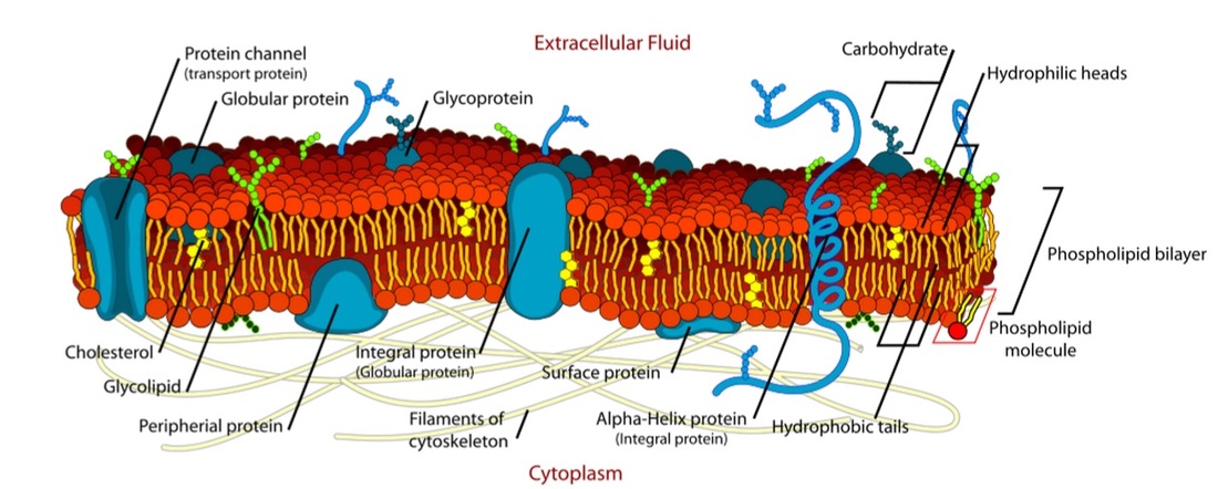
Cell Membrane:
Every cell is enclosed in a membrane, a double layer of phospholipids (lipid bilayer). The exposed heads of the bilayer are "hydrophilic" (water loving), meaning that they are compatible with water both within the cytosol and outside of the cell. However, the hidden tails of the phosopholipids are "hydrophobic" (water fearing), so the cell membrane acts as a protective barrier to the uncontrolled flow of water. The membrane is made more complex by the presence of numerous proteins that are crucial to cell activity. These proteins include receptors for odors, tastes and hormones, as well as pores responsible for the controlled entry and exit of ions like sodium (Na+) potassium (K+), calcium (Ca++) and chloride (Cl-).
Mitochondria:
Mitochondria provide the energy a cell needs to move, divide, produce secretory products, contract - in short, they are the power centers of the cell. They are about the size of bacteria but may have different shapes depending on the cell type. Mitochondria are membrane-bound organelles, and like the nucleus have a double membrane. The outer membrane is fairly smooth. But the inner membrane is highly convoluted, forming folds (cristae) as seen in the cross-section, above. The cristae greatly increase the inner membrane's surface area. It is on these cristae that food (sugar) is combined with oxygen to produce ATP - the primary energy source for the cell.
Vacuole:
A vacuole is a membrane-bound sac that plays roles in intracellular digestion and the release of cellular waste products. In animal cells, vacuoles are generally small. Vacuoles tend to be large in plant cells and play several roles: storing nutrients and waste products, helping increase cell size during growth, and even acting much like lysosomes of animal cells. The plant cell vacuole also regulates turgor pressure in the cell. Water collects in cell vacuoles, pressing outward against the cell wall and producing rigidity in the plant. Without sufficient water, turgor pressure drops and the plant wilts.
Cell Wall (plant cells only):
Plant cells have a rigid, protective cell wall made up of polysaccharides. In higher plant cells, that polysaccharide is usually cellulose. The cell wall provides and maintains the shape of these cells and serves as a protective barrier. Fluid collects in the plant cell vacuole and pushes out against the cell wall. This turgor pressure is responsible for the crispness of fresh vegetables.
Chloroplast (plant cells only):
Chloroplasts are specialized organelles found in all higher plant cells. These organelles contain the plant cell's chlorophyll responsible for the plant's green color and the ability to absorb energy from sunlight. This energy is used to convert water plus atmospheric carbon dioxide into metabolizable sugars by the biochemical process of photosynthesis. Chloroplasts have a double outer membrane. Within the stroma are other membrane structures - the thylakoids. Thylakoids appear in stacks called "grana" (singular = granum).
Smooth Endoplasmic Reticulum:
Throughout the eukaryotic cell, especially those responsible for the production of hormones and other secretory products, is a vast network of membrane-bound vesicles and tubules called the endoplasmic reticulum, or ER for short. The ER is a continuation of the outer nuclear membrane and its varied functions suggest the complexity of the eukaryotic cell.
The smooth endoplasmic reticulum is so named because it appears smooth by electron microscopy. Smooth ER plays different functions depending on the specific cell type including lipid and steroid hormone synthesis, breakdown of lipid-soluble toxins in liver cells, and control of calcium release in muscle cell contraction.
Rough Endoplasmic Reticulum:
Rough endoplasmic reticulum appears "pebbled" by electron microscopy due to the presence of numerous ribosomes on its surface. Proteins synthesized on these ribosomes collect in the endoplasmic reticulum for transport throughout the cell - it is a site of protein synthesis.
Ribosomes:
Ribosomes are packets of RNA and protein that play a crucial role in both prokaryotic and eukaryotic cells. They are the site of protein synthesis. Each ribosome comprises two parts, a large subunit and a small subunit. Messenger RNA from the cell nucleus is moved systematically along the ribosome where transfer RNA adds individual amino acid molecules to the lengthening protein chain.
Cytoskeleton:
As its name implies, the cytoskeleton helps to maintain cell shape. But the primary importance of the cytoskeleton is in cell motility. The internal movement of cell organelles, as well as cell locomotion and muscle fiber contraction could not take place without the cytoskeleton. The cytoskeleton is an organized network of three primary protein filaments:
- microtubules
- actin filaments (microfilaments)
- intermediate fibers
Advantages of eukaryotic cells being compartmentalized:
- Enzymes and substrates for a particular process can be more concentrated.
- Substances harmful to the cell can be contained within the membrane of an organelle. (e.g., digestive enzymes in lysosomes)
- Ideal conditions for a particular process can be maintained. (e.g. pH)
- Organelles with their contents can be moved around within the cell.
U1.2.3 Electron microscopes have a much higher resolution than light microscopes.
An electron microscope uses an electron beam to illuminate a specimen and produce the image.
An optical or light microscope uses a light beam to illuminate a specimen and produce the image.
A microscope of either type is characterized by its magnification and resolving power. The magnification depends on the lensing system and can be increased to any degree, but the maximum useful magnification is limited by the resolving power.
The resolving power of a microscope can not be better than the limits placed on it by the size of the wavelength of the illuminating beam. The smaller the wavelength, the smaller the structures that can be resolved in them image.
APPLICATION:
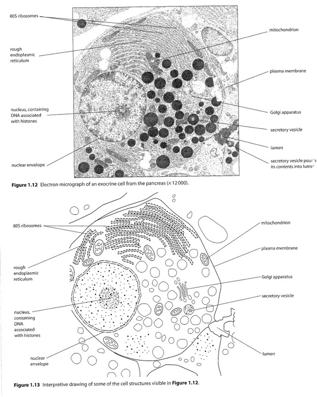
A1.2.1 Structure and function of organelles within exocrine gland cells of the pancreas and within palisade mesophyll cells of the leaf.
Exocrine gland cells in the pancreas are responsible for secreting digestive enzymes that travel to the small intestines where they aid in the digestion of food. Since these enzymes are proteins, exocrine gland cells have organelles that synthesize proteins, process them for secretion, transport them to the plasma membrane and then release them. These organelles include: nucleus, rough ER, Golgi apparatus, vesicles, lysosomes, and plasma membrane.
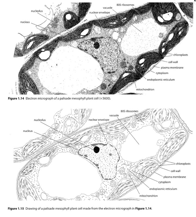
Most photosynthesis takes place in the palisade mesophyll cells of the leaf. Photosynthesis produces organic compounds (glucose) from carbon dioxide and simple inorganic compounds using light energy. The palisade mesophyll cells contain the following organelles: cell wall, plasma membrane, chloroplasts, mitochondrion, vacuole, and nucleus.
SKILL:
S1.2.1 Drawing of the ultrastructure of prokaryotic cells based on electron micrographs.
S1.2.2 Drawing of the ultrastructure of eukaryotic cells based on electron micrographs.
S1.2.1 Drawing of the ultrastructure of prokaryotic cells based on electron micrographs.
S1.2.2 Drawing of the ultrastructure of eukaryotic cells based on electron micrographs.
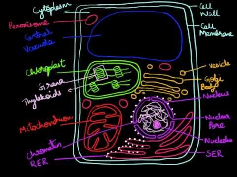
NOTE: there more than one type of eukaryotic cell - this drawing is of a plant cell.
S1.2.3 Interpretation of electron micrographs to identify organelles and deduce the function of specialized cells.
S1.2.3 Interpretation of electron micrographs to identify organelles and deduce the function of specialized cells.
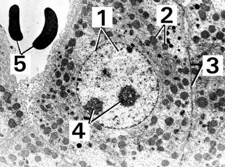
1. nucleus
2. mitochondria
3. plasma membrane
4. nucleoli
5. red blood cells (in adjacent blood vessel)
2. mitochondria
3. plasma membrane
4. nucleoli
5. red blood cells (in adjacent blood vessel)
Essential idea: The structure of biological membranes makes them fluid and dynamic.
1.3 Membrane structure
UNDERSTANDINGS:
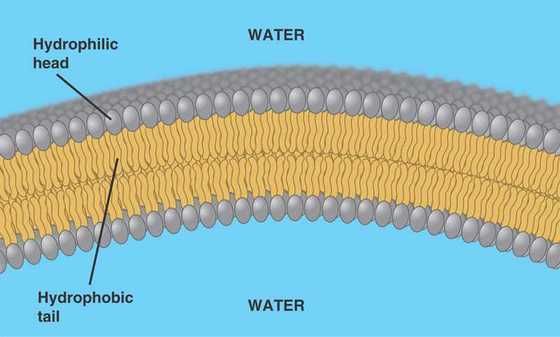
U1.3.1 Phospholipids form bilayers in water due to the amphipathic properties of phospholipid molecules.
The hydrophobic and hydrophilic regions cause phospholipids to always align as a bilayer if there is water present and there is a large number of phospholipid molecules. Because the fatty acid 'tails' do not strongly attract one another, the membrane tends to be fluid or flexible. This allows animal cells to have a variable shape and also allows the process of endocytosis. What maintains the overall structure of the membrane is the tendency water has to form hydrogen bonds.
Structural Properties of Phospholipid Bilayer
UNDERSTANDINGS:
U1.3.1 Phospholipids form bilayers in water due to the amphipathic properties of phospholipid molecules.
- Phospholipids consist of a glycerol molecule, two fatty acids, and a phosphate group that is modified by an alcohol.
- The phosphate group is the negatively-charged polar head, which is hydrophilic (i.e. attracts water).
- The fatty acid chains are the uncharged, nonpolar tails, which are hydrophobic (i.e. repels water).
- Since the tails are hydrophobic, they face the inside, away from the water and meet in the inner region of the membrane.
- Since the heads are hydrophilic, they face outward and are attracted to the intracellular and extracellular fluid.
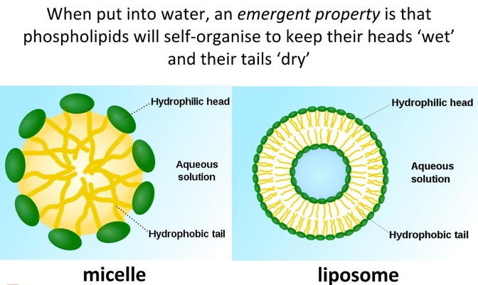
- If phospholipids are placed in water, they form into micelles, which are lipid molecules that arrange themselves in a spherical form in aqueous solutions.
The hydrophobic and hydrophilic regions cause phospholipids to always align as a bilayer if there is water present and there is a large number of phospholipid molecules. Because the fatty acid 'tails' do not strongly attract one another, the membrane tends to be fluid or flexible. This allows animal cells to have a variable shape and also allows the process of endocytosis. What maintains the overall structure of the membrane is the tendency water has to form hydrogen bonds.
Structural Properties of Phospholipid Bilayer
- Phospholipids are held together in a bilayer by hydrophobic interactions (weak associations)
- Hydrophilic / hydrophobic layers restrict entry and exit of substances
- Phospholipids allow for membrane fluidity / flexibility (important for functionality)
- Phospholipids with short or unsaturated fatty acids are more fluid
- Phospholipids can move horizontally or occasionally laterally to increase fluidity
- Fluidity allows for the breaking / remaking of membranes (exocytosis / endocytosis)
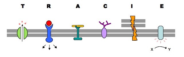
U1.3.2 Membrane proteins are diverse in terms of structure, position in the membrane and function.
Hormone binding sites (receptor proteins = glycoprotein)
- Proteins embedded in the membrane can bind to specific hormones.
- When the hormone binds, it causes the receptor protein to undergo a conformational change, which signals the cell to perform a function.
- For example, insulin receptors.
- Integral proteins that catalyse specific chemical reactions.
- Many of these enzymes catalyse metabolic reactions or are a part of a metabolic pathway, such as ATP Synthase in aerobic respiration.
- Proteins that form tight bonds between adjacent cells in tissues and organs.
- For example, gap junctions.
- Receptors for neurotransmitters at synapses between two nerve cells.
- Glycoproteins and peripheral proteins can also be used for cell identification purposes.
- Integral proteins that span the membrane and provide a passageway for molecules to move from an area of high concentration to low concentration.
- Specific proteins are also used for facilitated diffusion.
- Channel proteins are used for passive transport.
- Proteins that use ATP to move substances from a low concentration to a high concentration across the membrane.
- For example, Sodium/Potassium (Na+/K+) pumps and the proton (H+) pumps.
An easy was to remember these functions:
Transport: Protein channels (facilitated) and protein pumps (active)
Receptors: Peptide-based hormones (insulin, glucagon, etc.)
Anchorage: Cytoskeleton attachments and extracellular matrix
Cell recognition: MHC proteins and antigens
Intercellular joinings: Tight junctions and plasmodesmata
Enzymatic activity: Metabolic pathways (e.g. electron transport chain)
U1.3.3 Cholesterol is a component of animal cell membranes.
This is why marine organisms for example living in polar regions have a high proportion of cholesterol in their membranes - to prevent the membrane from become too inflexible.
APPLICATION:
A1.3.1 Cholesterol in mammalian membranes reduces membrane fluidity and permeability to some solutes.
SKILL:
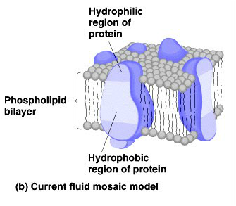
S1.3.1 Drawing of the fluid mosaic model.
- Cholesterol is required to build and maintain cell membranes - it regulates the permeability/fluidity of the membrane.
- Cholesterol is a lipid that belongs in the steroid group.
- Cholesterol is an amphipathic molecule, meaning, like phospholipids, it contains a hydrophilic and a hydrophobic portion. Cholesterol's hydroxyl (OH) group aligns with the phosphate heads of the phospholipids. The remaining portion of it tucks into the fatty acid portion of the membrane.
- Because of the way cholesterol is shaped, part of the steroid ring (the four hydrocarbon rings in between the hydroxyl group and the hydrocarbon "tail") is closely attracted to part of the fatty acid chain on the nearest phospholipid. This helps slightly immobilize the outer surface of the membrane and make it less soluble to very small water-soluble molecules that could otherwise pass through more easily.
This is why marine organisms for example living in polar regions have a high proportion of cholesterol in their membranes - to prevent the membrane from become too inflexible.
APPLICATION:
A1.3.1 Cholesterol in mammalian membranes reduces membrane fluidity and permeability to some solutes.
SKILL:
S1.3.1 Drawing of the fluid mosaic model.
- Phospholipid bilayer annotated to show composition of phosphate heads and fatty acid tails
- Cholesterol shown between phospholipids, stretching inward from phospholipid surface along side fatty acid tails
- Glycoproteins shown with carbohydrate chain attached to integral protein and extending outside cell membrane
- Integral proteins embedded within the phospholipids of the membrane
- Transmembrane proteins extending fully across phospholipid membrane
- Peripheral proteins attached to the outside of the phospholipid surface
- Glycolipid shown with carbohydrate chain attached phospholipid head and extending outside cell membrane
- Annotated to show membrane thickness of 10 nm
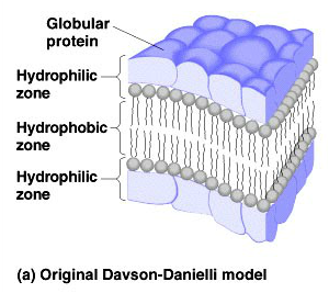
S1.3.2 Analysis of evidence from electron microscopy that led to the proposal of the Davson-Danielli model.
The Davson-Danielli model of the plasma membrane was the working model from 1935-1972. Scientists knew that the membrane was composed of lipids largely due to the fact that fat soluble substances easily dissolved in the membrane. This model consisted of a phospholipid interior surrounded by protein coats on each side.
In high magnification electron micrograph membranes appeared as 2 dark parallel lines with a lighter coloured region in between. Proteins appears dark in electron micrographs and phospholipids appear light - possibly indicating protein layers either side of a phospholipid core.
Scientists knew that the membrane was composed of lipids largely due to the fact that fat soluble substances easily dissolved in the membrane.
The model:
1. A protein-lipid sandwich
2. Lipid bilayer composed of phospholipids (hydrophobic tails inside and hydrophilic heads outside)
3. Proteins coats outer surface
4. Proteins do not permeate the lipid bilayer
S1.3.3 Analysis of the falsification of the Davson-Danielli model that led to the Singer-Nicolson model.
While the arrangement of the phospholipids were correct, there was issue with the arrangement of the proteins. They are also amphipathic and they would be blocking the hydrophilic heads of the phospholipids from the aqueous external environment. The model also did not allow for the different structures and functions of membrane proteins.
The Singer-Nicholson model was proposed in 1972 and remains the current accepted model of the plasma membrane. It is known as the fluid mosaic model and while the arrangements of the phospholipids remain the same, it is the location of the proteins that differs. This model shows them embedded in the bilayer as either integral or peripheral proteins.
Essential idea: Membranes control the composition of cells by active and passive transport.
1.4 Membrane transport
U1.4.1 Particles move across membranes by simple diffusion, facilitated diffusion, osmosis and active transport.
Membranes are semi-permeable which means that they allow certain molecules through but not others. The molecules can move in and out through passive transport which is a method that does not require any input of outside energy. It can either be done by simple diffusion or facilitated diffusion.
Molecules will go from a region of high concentration to a region of low concentration as they move randomly and eventually become evenly distributed within the system if they are permeable to the membrane. Simple diffusion involves the diffusion of molecules through the phospholipid bilayer while facilitated diffusion involves the use of channel proteins embedded in the membrane.
The cell membrane is hydrophobic inside so hydrophobic (lipid soluble) molecules will pass through by simple diffusion whereas hydrophilic molecules and charged particles will use facilitated diffusion. Water moves through by osmosis which is also by passive transport. Osmosis involves the movement of water molecules from a region of low solute concentration, to a region of high solute concentration. So if the solute concentration is higher inside the cell than outside the cell, water will move in and vice versa.
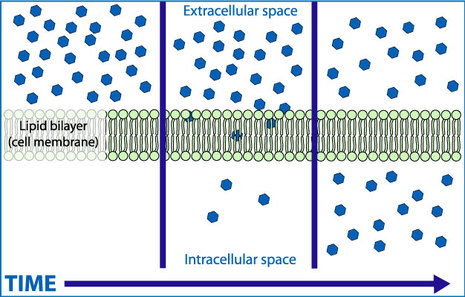
Simple diffusion:
In simple diffusion, small non-charged molecules or lipid soluble molecules pass between the phospholipids to enter or leave the cell, moving from areas of high concentration to areas of low concentration (they move down their concentration gradient). Oxygen and carbon dioxide and most lipids enter and leave cells by simple diffusion.
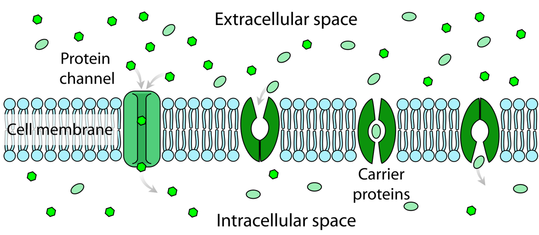
Facilitated diffusion:
In facilitated diffusion, substances move into or out of cells down their concentration gradient through protein channels in the cell membrane. Simple diffusion and facilitated diffusion are similar in that both involve movement down the concentration gradient. The difference is how the substance gets through the cell membrane. In simple diffusion, the substance passes between the phospholipids; in facilitated diffusion there are a specialised membrane channels. Charged or polar molecules that cannot fit between the phospholipids generally enter and leave cells through facilitated diffusion.
A special carrier protein with a central channel (integral proteins) acts as a selective corridor which helps molecules move across the membrane. Each carrier protein is designed to recognise only one substance or one group of very similar substances i.e. specific molecules such as a particular sugar or amino acid will only bind to a specific protein. Once the molecule binds to the carrier protein, this protein facilitates the diffusion process by changing shape and moving the molecule down its concentration gradient through the membrane into the cell where it is released. Facilitated diffusion can happen in either direction - if there is a higher concentration of the particular molecule inside the cell, the same carrier protein will transport the molecules out of the cell.
specific molecule such as a particular sugar or amino acid.
U1.4.1 Particles move across membranes by simple diffusion, facilitated diffusion, osmosis and active transport.
Membranes are semi-permeable which means that they allow certain molecules through but not others. The molecules can move in and out through passive transport which is a method that does not require any input of outside energy. It can either be done by simple diffusion or facilitated diffusion.
Molecules will go from a region of high concentration to a region of low concentration as they move randomly and eventually become evenly distributed within the system if they are permeable to the membrane. Simple diffusion involves the diffusion of molecules through the phospholipid bilayer while facilitated diffusion involves the use of channel proteins embedded in the membrane.
The cell membrane is hydrophobic inside so hydrophobic (lipid soluble) molecules will pass through by simple diffusion whereas hydrophilic molecules and charged particles will use facilitated diffusion. Water moves through by osmosis which is also by passive transport. Osmosis involves the movement of water molecules from a region of low solute concentration, to a region of high solute concentration. So if the solute concentration is higher inside the cell than outside the cell, water will move in and vice versa.
Simple diffusion:
In simple diffusion, small non-charged molecules or lipid soluble molecules pass between the phospholipids to enter or leave the cell, moving from areas of high concentration to areas of low concentration (they move down their concentration gradient). Oxygen and carbon dioxide and most lipids enter and leave cells by simple diffusion.
Facilitated diffusion:
In facilitated diffusion, substances move into or out of cells down their concentration gradient through protein channels in the cell membrane. Simple diffusion and facilitated diffusion are similar in that both involve movement down the concentration gradient. The difference is how the substance gets through the cell membrane. In simple diffusion, the substance passes between the phospholipids; in facilitated diffusion there are a specialised membrane channels. Charged or polar molecules that cannot fit between the phospholipids generally enter and leave cells through facilitated diffusion.
A special carrier protein with a central channel (integral proteins) acts as a selective corridor which helps molecules move across the membrane. Each carrier protein is designed to recognise only one substance or one group of very similar substances i.e. specific molecules such as a particular sugar or amino acid will only bind to a specific protein. Once the molecule binds to the carrier protein, this protein facilitates the diffusion process by changing shape and moving the molecule down its concentration gradient through the membrane into the cell where it is released. Facilitated diffusion can happen in either direction - if there is a higher concentration of the particular molecule inside the cell, the same carrier protein will transport the molecules out of the cell.
specific molecule such as a particular sugar or amino acid.
Osmosis:
Osmosis is a special example of diffusion. It is the passive movement of water molecules, across a partially permeable membrane, from a region of lower solute concentration to a region of higher solute concentration.
NOTE: diffusion and osmosis are both passive, i.e. energy from ATP is not used.
A partially permeable membrane is a barrier that permits the passage of some substances but not others; it allows the passage of the solvent molecules but not some of the larger solute molecules. Cell membranes are described as selectively permeable because not only do they allow the passage of water but also allow the passage of certain solutes. The presence of particular solutes stimulates the membrane to open specific channels or trigger active transport mechanisms to allow the passage of those chemicals across the membrane.
Some major examples of osmosis:
• Absorption of water by plant roots
• Re-absorption of water by the proximal and distal convoluted tubules of the nephron
• Re-absorption of tissue fluid into the venule ends of the blood capillaries
• Absorption of water by the alimentary canal — stomach, small intestine and the colon.
Active transport:
Active transport involves the movement of substances through the membrane using energy from ATP. The advantage of active transport is that substances can be moved against the concentration gradient, meaning from a region of low concentration to a region of high concentration. This is possible because the cell membrane has protein pumps embedded it which are used in active transport to move substances across by using ATP. Each protein pump only transports certain substances so the cell can control what comes in and what goes out.
The sodium potassium (NaK+) pump is an active transport mechanism. 3 sodium ions bind to the protein channel and an ATP molecule provides the energy required to change the shape of the channel that in turn drives the ions through the channel. 1 phosphate group from the ATP remains bound with the protein channel. The sodium ions are released on the other side of the membrane outside the cell and the new shape of the channel has a high affinity for potassium ions and 2 of these ions now bind to the channel. This binding again causes a change in the shape of the protein channel and this conformational change releases the phosphate group on the cytoplasm side. This release allows the channel to revert to its original shape and as a result, the potassium ions are released inside the cell.
In its original shape, the protein channel has a high affinity for sodium ions and when these ions bind again, they initiate another cycle. The important characteristic of this pump is that both the sodium and potassium ions are moving from regions of low concentration to regions of high concentration i.e. each ion is moving against its concentration gradient. This movement can only be achieved by the constant expenditure of ATP energy.
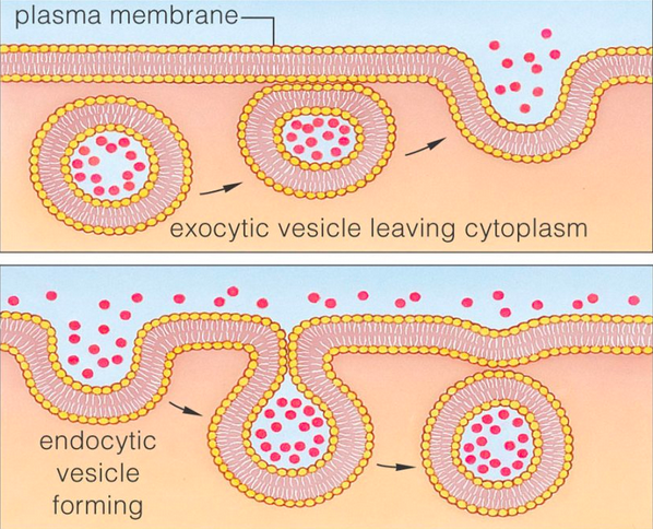
U1.4.2 The fluidity of membranes allows materials to be taken into cells by endocytosis or released by exocytosis. Vesicles move materials within cells.
Endocytosis: The taking in of external substances by an inward pouching of the plasma membrane, forming a vesicle
Exocytosis: The release of substances from a cell (secretion) when a vesicle joins with the cell plasma membrane.
Factors affecting fluidity:
1. Temperature
2. Cholesterol
3. Un/saturated fatty acids
1. Low temperature = phospholipids cluster together and do not move around a lot due to low levels of kinetic energy = low fluidity
High temperature = increased distance between phospholipids = high fluidity
2. Effect of cholesterol at low temperatures = increases distance between phospholipids = higher fluidity
Effect of cholesterol at high temperatures = causes phospholipids to pull themselves closer together because they want to 'attach' to the cholesterol = lower fluidity!
Cholesterol regulates fluidity - it acts as a buffer.
3. Saturated fats contain only single bonds however unsaturated fats contain double bonds.
Saturated fats stack evenly like Legos but unsaturated fats do not.
Since sat. fats stack nicely, the phospholipids sit closely together = low fluidity
Since unsat. fats contain double bonds i.e. 'bends' in the bonds, the phospholipids are not able to sit as closely together = high fluidity
Endocytosis:
- Plasma membrane is being pinched as a result of the membrane changing shape
- External materials i.e. fluid droplets are engulfed and enclosed by the membrane
- A vesicle is formed that contain the enclosed particles now moves into the cytoplasm
- The plasma membrane easily reattaches at the ends that were pinched because of the fluidity of the membrane
- Vesicles that move through the cytoplasm are broken down and dissolve into the cytoplasm.
NOTE: In cell biology, a vesicle is a small organelle within a cell, consisting of fluid enclosed by a lipid bilayer membrane. Vesicles can form naturally, for example, during the processes of secretion (exocytosis), uptake (phagocytosis and endocytosis) and transport of materials within the cytoplasm.
Exocytosis:
- After a vesicle created by the rough ER enters the Golgi apparatus, it is again modified and another vesicle is budded from the end of the Golgi, which moves towards the cell membrane
- The vesicle migrates to the plasma membrane and fuses with the membrane, releasing the particle outside the cell through a process called exocytosis.
APPLICATION:
A1.4.1 Structure and function of sodium–potassium pumps for active transport and potassium channels for facilitated diffusion in axons.
- Axon – part of a neurone (nerve cell) and consists of a tubular membrane with cytoplasm inside - can be as narrow as one micrometer in diameter but as long as one meter
- Their jobs is to convey messages in an electrical form called a nerve impulse
- Nerve impulse is rapid movements of sodium and then potassium ions across the axon membrane - these movements occur by facilitated diffusion through sodium and potassium channels and this occurs because of concentration gradients between the inside and outside of the axon
- The concentration gradients are built up by active transport, carried out by a sodium potassium pump protein
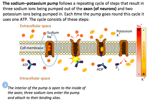
- The sodium-potassium pump follows a repeating cycle of steps that result in three sodium ions being pumped out of the axon and two potassium ions being pumped in
- This process uses one ATP molecule
- The inside of the pump is open to the inside of the axon; three sodium ions enter the pump and attach to their binding sites
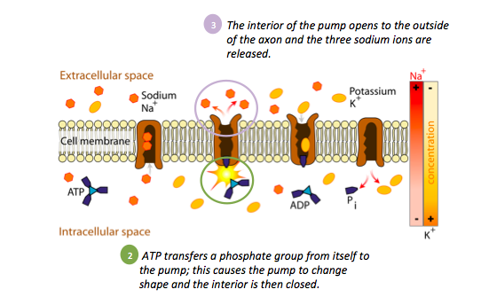
- ATP transfers a phosphate group from itself to the pump; this causes the pump to change shape and the interior is then closed
- The inside of the pump opens to the outside of the axon and the three sodium ions are released
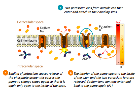
- Two potassium ions from outside can then enter and attach to their binding sites
- Binding of potassium = release of the phosphate group; this causes the pump to change shape again so that it is again only open to the of the axon
- The inside of the pump opens to the inside of the axon and the two potassium ions are released; sodium ions can then enter and bind to the pump again
A1.4.2 Tissues or organs to be used in medical procedures must be bathed in a solution with the same osmolarity as the cytoplasm to prevent osmosis.
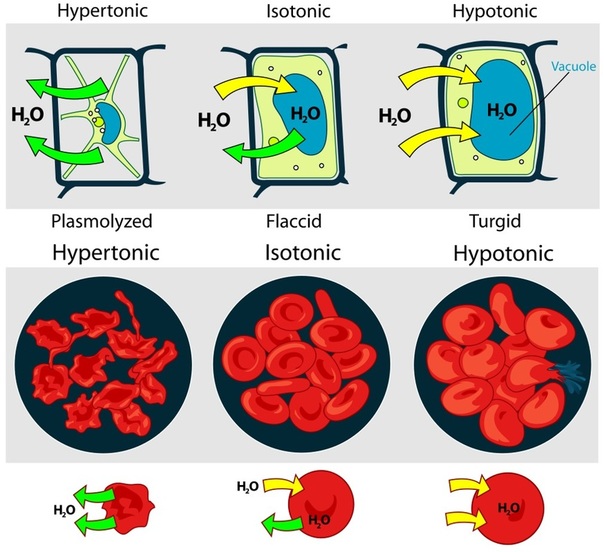
Hypertonic solution is a solution with a higher osmolarity (higher solute concentration) then the other solution. If cells are placed into a hypertonic solution, water will leave the cell causing the cytoplasm’s volume to shrink and thereby forming indentations in the cell membrane.
Hypotonic solution is a solution with a lower osmolarity (lower solute concentration) then the other solution. If cells are placed in a hypotonic solution, the water will rush into the cell causing them to swell and possibly burst.
Both of the above solutions would damage cells, therefore isotonic solutions are used (same osmolarity as inside the cell). An isotonic solution is a solution that has the same salt concentration as cells and blood.
Kidney dialysis:
Dialysis is the artificial process of eliminating waste (diffusion) and unwanted water (ultrafiltration) from the blood. Our kidneys do this naturally. Some people, however, may have failed or damaged kidneys which cannot carry out the function properly - they may need dialysis. In other words, dialysis is the artificial replacement for lost kidney function (renal replacement therapy). Dialysis may be used for patients who have become ill and have acute kidney failure (temporary loss of kidney function), or for fairly stable patients who have permanently lost kidney function.
Kidney dialysis is a life-support treatment that uses a special machine to filter the harmful wastes, salt and excess fluid from your blood. This restores the blood to a normal, healthy balance. Dialysis replaces many of the kidney's important functions.
There are different types of kidney dialysis, including:
Before hemodialysis can be done, a doctor must make an entrance, called an access, into the patient's blood vessels. This is done by minor surgery in the leg, arm or sometimes neck. The best access for most patients is called a fistula. Minor surgery is performed to join an artery to a vein under the skin to make a larger vessel.
Blood drains into the dialysis machine to be cleaned. The machine has two parts, one side for blood and one for a fluid called dialysate. A thin, semipermeable membrane separates the two parts. As dialysate passes on one side of the membrane, and blood on the other, particles of waste from the blood pass through microscopic holes in the membrane and are washed away in the dialysate. Blood cells are too large to go through the membrane and are returned to the body.
Hypertonic solution is a solution with a higher osmolarity (higher solute concentration) then the other solution. If cells are placed into a hypertonic solution, water will leave the cell causing the cytoplasm’s volume to shrink and thereby forming indentations in the cell membrane.
Hypotonic solution is a solution with a lower osmolarity (lower solute concentration) then the other solution. If cells are placed in a hypotonic solution, the water will rush into the cell causing them to swell and possibly burst.
Both of the above solutions would damage cells, therefore isotonic solutions are used (same osmolarity as inside the cell). An isotonic solution is a solution that has the same salt concentration as cells and blood.
Kidney dialysis:
Dialysis is the artificial process of eliminating waste (diffusion) and unwanted water (ultrafiltration) from the blood. Our kidneys do this naturally. Some people, however, may have failed or damaged kidneys which cannot carry out the function properly - they may need dialysis. In other words, dialysis is the artificial replacement for lost kidney function (renal replacement therapy). Dialysis may be used for patients who have become ill and have acute kidney failure (temporary loss of kidney function), or for fairly stable patients who have permanently lost kidney function.
Kidney dialysis is a life-support treatment that uses a special machine to filter the harmful wastes, salt and excess fluid from your blood. This restores the blood to a normal, healthy balance. Dialysis replaces many of the kidney's important functions.
There are different types of kidney dialysis, including:
- Hemodialysis. Blood is filtered using a dialyzer and dialysis machine.
- Peritoneal dialysis. Blood is filtered inside the body after the abdomen is filled with a special cleaning solution.
Before hemodialysis can be done, a doctor must make an entrance, called an access, into the patient's blood vessels. This is done by minor surgery in the leg, arm or sometimes neck. The best access for most patients is called a fistula. Minor surgery is performed to join an artery to a vein under the skin to make a larger vessel.
Blood drains into the dialysis machine to be cleaned. The machine has two parts, one side for blood and one for a fluid called dialysate. A thin, semipermeable membrane separates the two parts. As dialysate passes on one side of the membrane, and blood on the other, particles of waste from the blood pass through microscopic holes in the membrane and are washed away in the dialysate. Blood cells are too large to go through the membrane and are returned to the body.
S1.4.1 Estimation of osmolarity in tissues by bathing samples in
hypotonic and hypertonic solutions.
-
Osmosis experiments are a useful opportunity to stress the need for accurate mass and volume measurements in scientific experiments.
-
Osmosis experiments are a useful opportunity to stress the need for accurate mass and volume measurements in scientific experiments.
Essential idea: There is an unbroken chain of life from the first cells on Earth to all cells in organisms alive today.
1.5 The origin of cells
U1.5.1 Cells can only be formed by division of pre-existing cells.
U1.5.2 The first cells must have arisen from non-living material.
1) Production of carbon compounds such amino acids and sugars. Miler and Urey’s experiment showed how this could happen by passing water vapour through Ammonia, methane and hydrogen (early earth atmosphere). They added electricity to simulate lightening discharge. They found they could create amino acids and carbon compounds
2) Assembly of carbon compounds into polymers might have occurred at the deep sea hydrothermal vents, which could have supplied the inorganic compounds such as iron sulphide and thermal energy for the assembly
3) Formation of membranes would be possible if phospholipids were some of the first polymers created. These phospholipids would naturally form vesicles allowing for a different environment to exist inside compared to the surrounding water
4) Development of a mechanism for inheritance would be needed in order for the organism to replicate and pass its DNA on to the next generation. Current organisms need enzymes to replicate DNA; however, enzymes are created by the genes on the DNA. A possible solution to this would be RNA being the first nucleic acid formed because it is self-replicating and can also act as a catalyst.
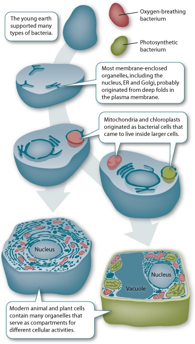
U1.5.3 The origin of eukaryotic cells can be explained by the endosymbiotic theory.
There is compelling evidence that mitochondria and chloroplasts were once primitive free-living bacterial cells.
U1.5.1 Cells can only be formed by division of pre-existing cells.
- Prokaryotic cells are formed during a process called binary fission (see A1.2.2).
- Eukaryotic cells form new identical cells by the process called mitosis (genetically identical) and form sex cells through meiosis (haploid cells which are not genetically identical to the parent cell and contain half the genetic material).
- Cell division to form new cells from pre-existing cells replaced the concept of spontaneous generation, where cells were formed from inanimate matter.
U1.5.2 The first cells must have arisen from non-living material.
- Abiogenesis is the natural process of life arising from non-living matter such as simple organic compounds
- If we go back to how the very first living cells were created, we have to conclude they either originated from non-living material, came from somewhere else in the universe or were created by some other unknown entity
- These are the hypothesized steps of how living cells possibly developed from non-living material over millions of years
1) Production of carbon compounds such amino acids and sugars. Miler and Urey’s experiment showed how this could happen by passing water vapour through Ammonia, methane and hydrogen (early earth atmosphere). They added electricity to simulate lightening discharge. They found they could create amino acids and carbon compounds
2) Assembly of carbon compounds into polymers might have occurred at the deep sea hydrothermal vents, which could have supplied the inorganic compounds such as iron sulphide and thermal energy for the assembly
3) Formation of membranes would be possible if phospholipids were some of the first polymers created. These phospholipids would naturally form vesicles allowing for a different environment to exist inside compared to the surrounding water
4) Development of a mechanism for inheritance would be needed in order for the organism to replicate and pass its DNA on to the next generation. Current organisms need enzymes to replicate DNA; however, enzymes are created by the genes on the DNA. A possible solution to this would be RNA being the first nucleic acid formed because it is self-replicating and can also act as a catalyst.
U1.5.3 The origin of eukaryotic cells can be explained by the endosymbiotic theory.
There is compelling evidence that mitochondria and chloroplasts were once primitive free-living bacterial cells.
- Symbiosis occurs when two different species benefit from living and working together. When one organism actually lives inside the other it's called endosymbiosis.
- The endosymbiotic theory describes how a large host cell and the bacteria ingested through endocytosis, could easily become dependent on one another for survival, resulting in a permanent relationship.
- As long as the smaller mitochondria living inside the cytoplasm of the larger cell divided at the same rate, they could persist indefinitely inside those cells
- The smaller cell was provided food and protection by the larger cell and the smaller mitochondria would supply energy through aerobic respiration for the larger cell
- Over millions of years of evolution, mitochondria and chloroplasts have become more specialized and today they cannot live outside the cell.